++ 50 ++ gram stain e coli under microscope 100x 186161
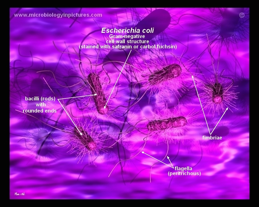
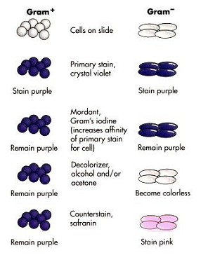
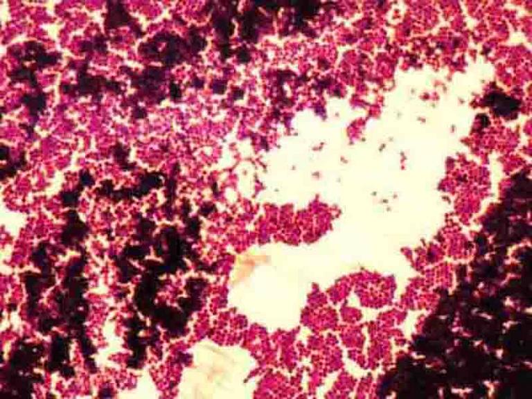
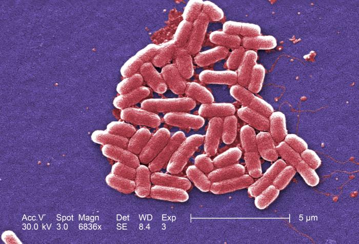
Biology Of E Coli E coli (Escherichia coli) are a small, Gramnegative species of bacteriaMost strains of E coli are rodshaped and measure about μm long and 0210 μm in diameterThey typically have a cell volume of 0607 μm, most of which is filled by the cytoplasm Since it is a prokaryote, E coli don't have nuclei;Characterizing E coli Using a Light Microscope and Gram Staining" from Biotechnology Laboratory Manual by Ellyn Daugherty For more and in sharp focus on 100X before going to 400X, and center and focused on 400X before adding the immersion E coli cells are GramThe slide is stained with safranin for 1min, then rinse with water very gently until no color is coming from the water Blot dry with absorbent paper Place the slide on the microscope to view the sample The grampositive bacteria will stain dark blue or purple The gramnegative bacteria will stain pink In this figure A procedure of Gram stain
Photomicrographs
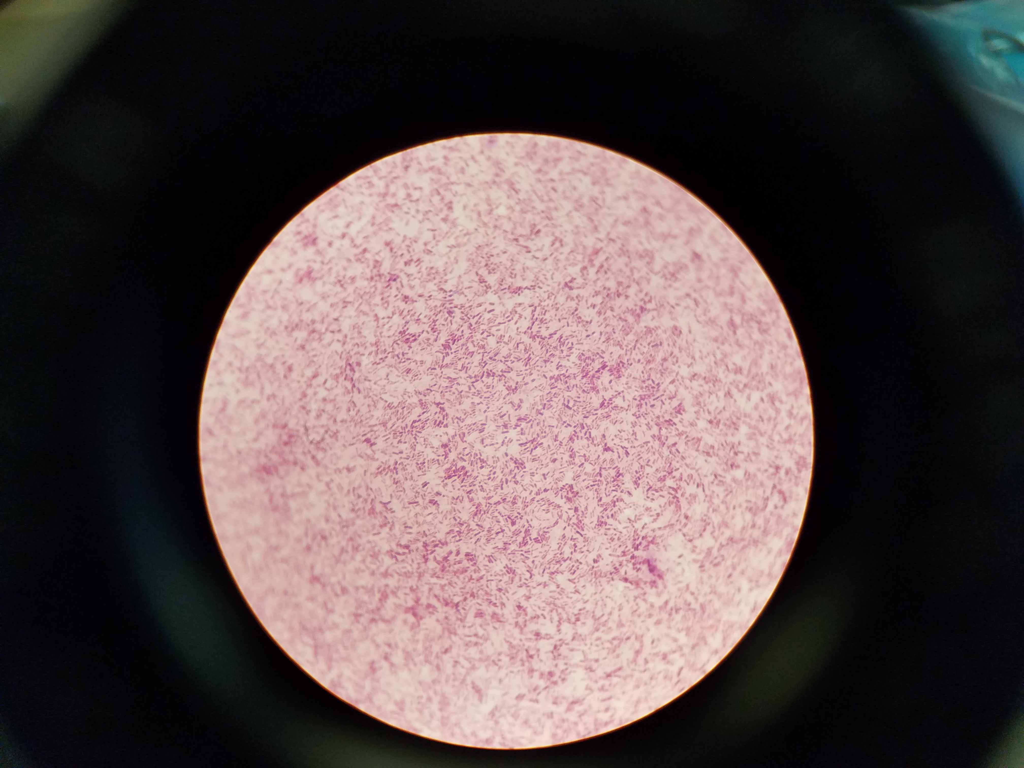
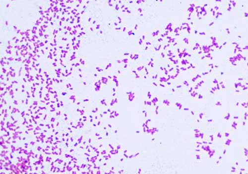
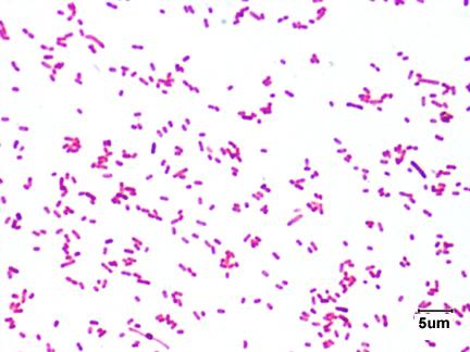
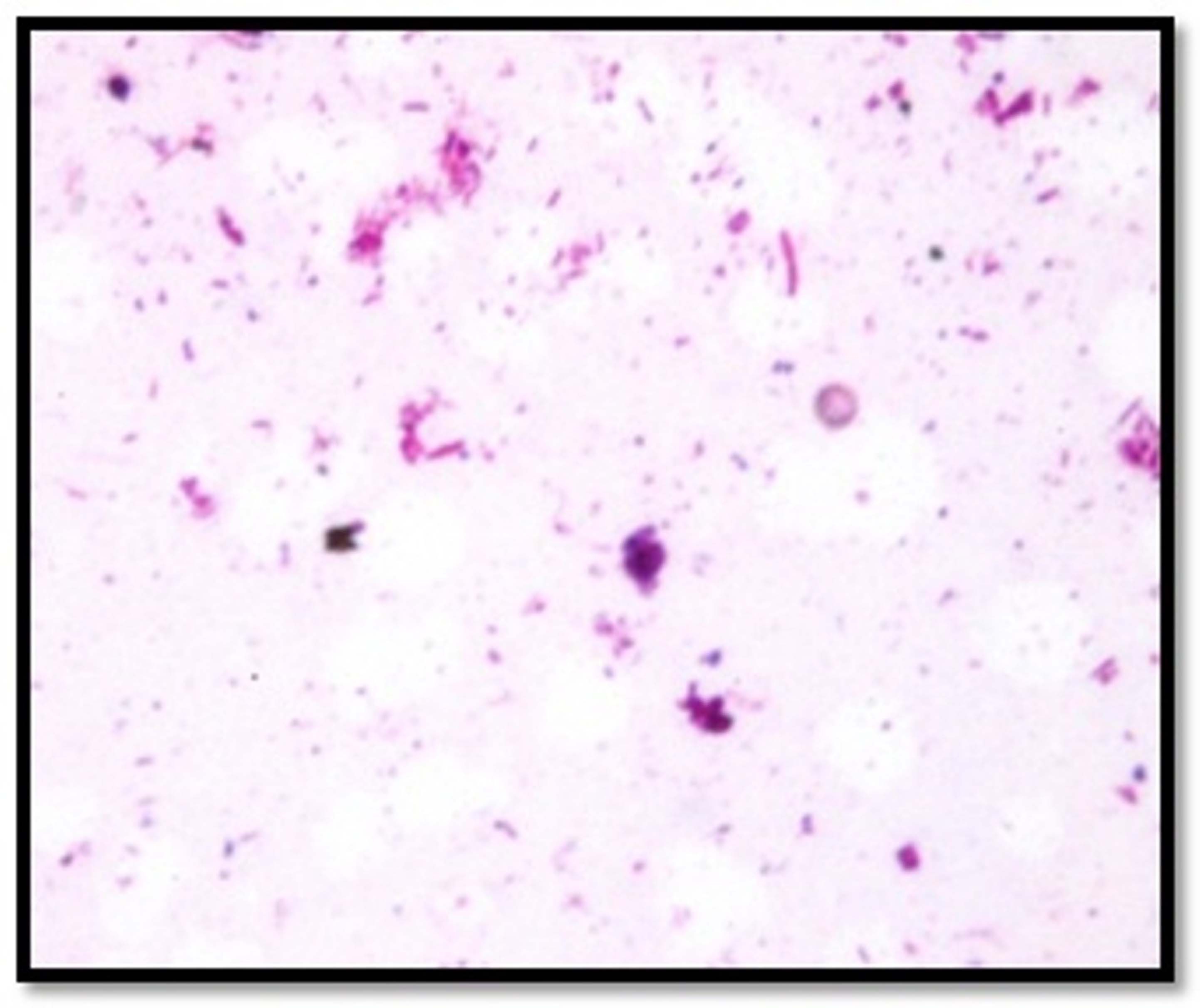
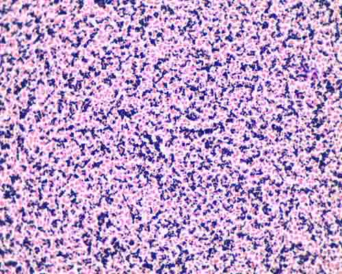
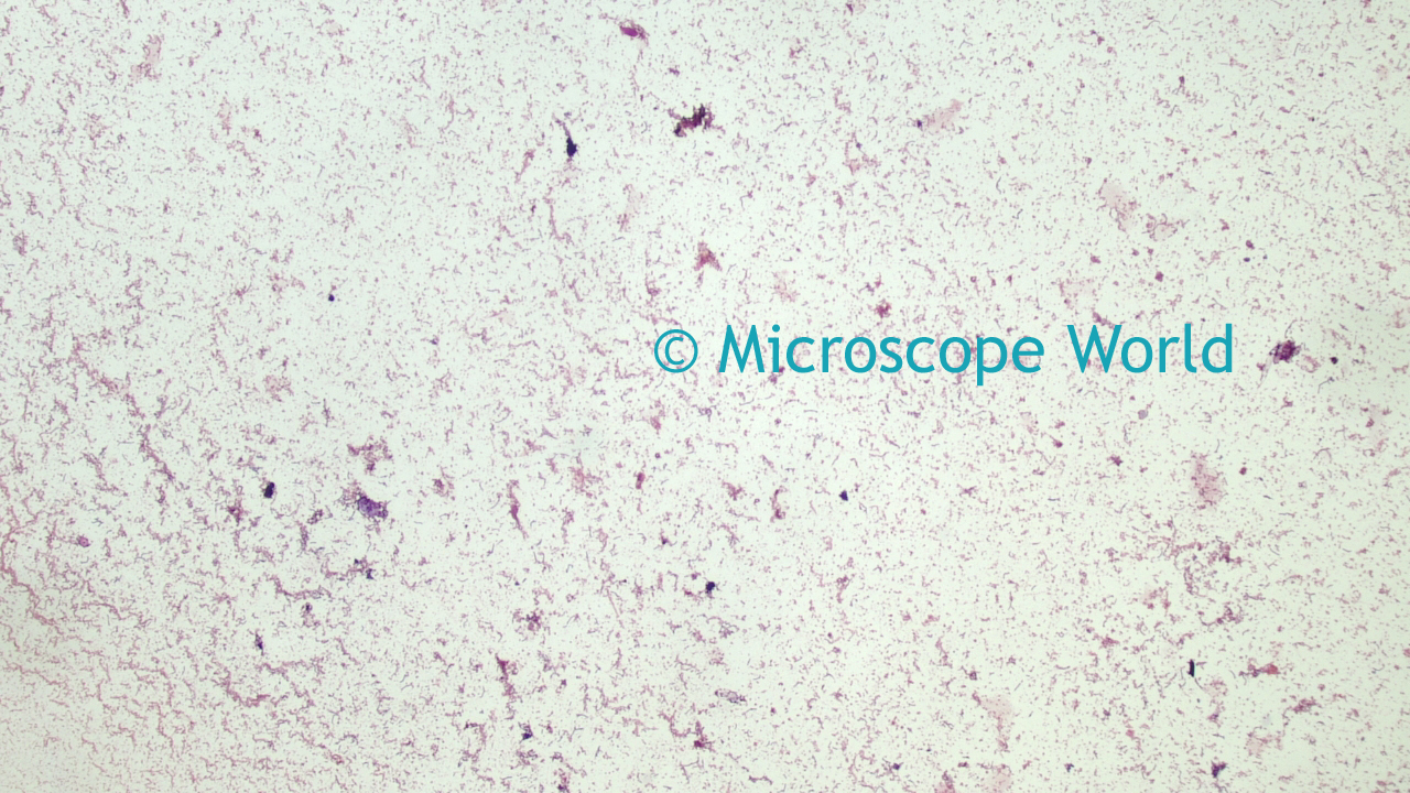
Gram stain e coli under microscope 100x
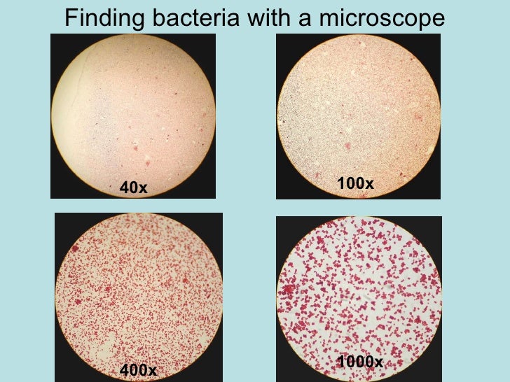
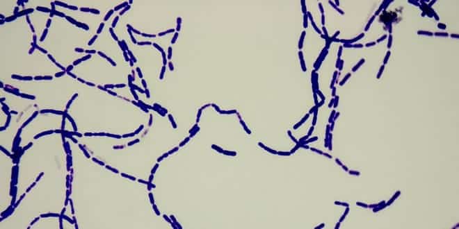
Gram stain e coli under microscope 100x-In contrast, Escherichia coli was rodshaped and pinkish red, it is Gramnegative fD) Structural Stains Endospore Stain Endospore Bacillus subtilis Figure 4 Bacillus subtilis and its endospores under the microscope with magnification of 100X The endospores were green in colour and Bacillus subtilis was red in colour1 E coli stained with crystal violet @ 100xTM It is not possible to see individual bacteria at this magnification, but viewing at 100XTM is a necessary step in focusing to scope to see at higher magnifications 2 E coli stained with crystal violet @ 1000xTM


Q Tbn And9gcqkye60ou Johpr02n Mbv1fferrjpdh Lnct7ymdf5qhyia1ld Usqp Cau
Tilt the slide slightly and gently rinse with tap water or distilled water using a wash bottle The smear will appear as a purple circle on the slide Decolorize using 95% ethyl alcohol or acetone Tilt the slide slightly and apply the alcohol drop by drop for 5 to 10 seconds until the alcohol runs almost clear1 E coli stained with crystal violet @ 100xTM It is not possible to see individual bacteria at this magnification, but viewing at 100XTM is a necessary step in focusing to scope to see at higher magnifications 2 E coli stained with crystal violet @ 1000xTMStain the slide with the primary stain, crystal violet Treat the stain with iodine (a mordant) Gently apply a decolorizer on the smear (s), then counterstain with safranin Grampositive microorganisms such as Staphylococcus aureus appear bluishpurple
Gram Staining of Micrococcus luteus,Escherichia coli, and Serratia marcescens INTRODUCTION In 14, a Danish botanist named Hans Christian Gram developed the popular staining technique known as the "gram stain"His background in studying plants at the University of Copenhagen introduced him to observing different tissues under a microscope, and thus propelled his interest in pharmacologyDIFFERENTIAL STAIN An example is Gram staining (or Gram's method) It is routinely used as an initial procedure in the identification of an unknown bacterial species Let's suppose we have a smear containing mixture of Staphylococcus aureus and Escherichia coli as in previous case We will use the same stains as before and besides we will needThere will not be any E Coli unless the plate was contaminated while it was left open on the bench top In viewing the simple stain of your mixed broth culture under 100x with oil, you will be able to describe the morphology of the cells present You observed a stained slide under the microscope, and found some cells were random and rod
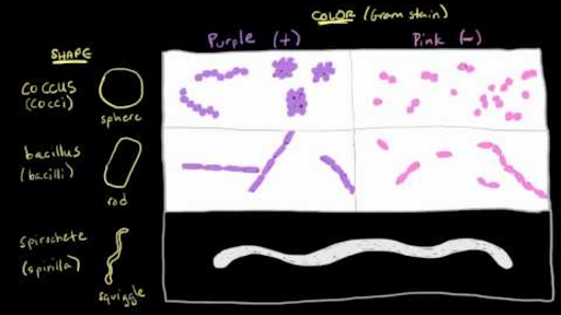
This could be because of E coli being a Gramnegative cell and therefore few cells decolorized when alcohol used, leading to pinkish color being visible when stained with safranin This is consistent with published results 1 We were successfully able to view the B subtilis cells using the 100x objective lensDuring the staining procedure, the Grampositive bacteria retain crystal violet and appear blue In contrast, Gramnegative lose crystal violet during the washing steps and hold only safranin to appear red Bacterial species Bacillus and Escherichia are the example of the Grampositive and Gramnegative respectively After Gram staining, the morphological difference between a coccid and rodshaped becomes much clearer under a compound light microscope with only 100X magnificationGram stain allows you to classify into Gramnegative rods (which includes E coli) and Grampositive rods (which include Bacillus, Corynebacterium, and some others)


Q Tbn And9gcqkye60ou Johpr02n Mbv1fferrjpdh Lnct7ymdf5qhyia1ld Usqp Cau
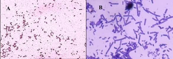


How These 26 Things Look Like Under The Microscope With Diagrams
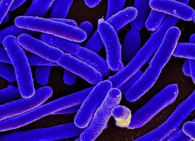
If you were performing a Gram stain and accidentally performed the decolorization step using water rather than 95% ethanol, what would your negative control look like (under the microscope, after completion of the entire GramStain procedure)?Gram stain e coli under microscope 100x › microscope e coli gram stain bacteria › microscope escherichia coli gram stain e coli Gram Stain E Coli Microscope Written By MacPride Tuesday, July 23, 19 Add Comment Edit Puzzlepix Figure 1 From Detection Of Multidrug Resistant And Shiga ToxinEscherichia coli or Ecoli is a gramnegative species of bacillus shaped bacteria that can be easily observed under a microscope, even for those with the untrained eye This bacteria has a fast growth rate, doubling every minutes, making them a common choice for bacterial related research purposes


Gram Stain


A Long Chain Like Colony Of Streptococcus Bacteria As Seen Under Download Scientific Diagram
Wash slide in a gentile and indirect stream of tap water until no color appears in the effluent and then blot dry with absorbent paper Observe the results of the staining procedure under oil immersion (100x) using a Bright field microscopeTHE PROCEDURE done individually We have cultures of E coli and Bacillus for you to gram stainThis will give you gram and gram – controls to check your procedure against You can use 2 slides, 1 for each bacterium, or you can divide one slide in half and smear each bacterium on the divided slideThe slide is stained with safranin for 1min, then rinse with water very gently until no color is coming from the water Blot dry with absorbent paper Place the slide on the microscope to view the sample The grampositive bacteria will stain dark blue or purple The gramnegative bacteria will stain pink In this figure A procedure of Gram stain


Solved 3 A Mixed Smear Of A 3 Day 72 Hour Culture Of S Chegg Com
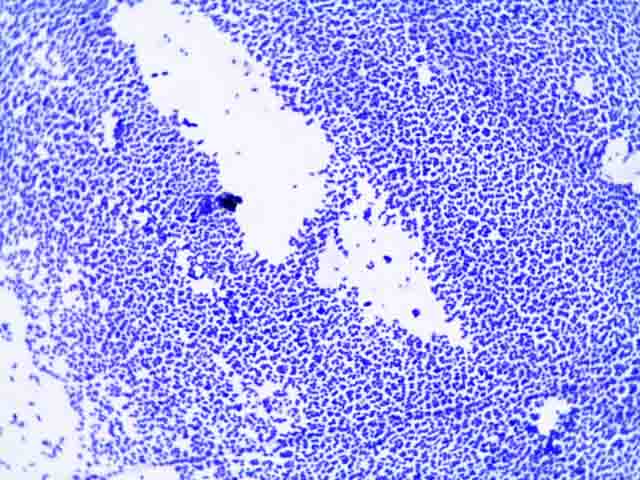


Bacterial Staining Microbiology Images Photographs And Videos Of Gram Acid Fast Endospore
Gram stain e coli under microscope 100x › microscope e coli gram stain bacteria › microscope escherichia coli gram stain e coli Gram Stain E Coli Microscope Written By MacPride Tuesday, July 23, 19 Add Comment Edit Puzzlepix Figure 1 From Detection Of Multidrug Resistant And Shiga ToxinWhen viewing a slide under 100X oil immersion, the diaphragm should be fully opened, allowing in the most light possible A fellow student is unsure why his gramnegative Escherichia coli is purple Upon examining the Gram stain kit, you notice it didn't have alcohol which prevented the Gram stain's iodine and crystal violet from beingE Coli (Escherichia Coli) is a gramnegative, rodshaped bacterium Most E Coli strains are harmless, but some serotypes can cause food poisoning in their hosts The harmless strains are part of the normal flora of the gut Learn more about E Coli here Helicobacter Pylori The prepared microscope slide image of Helicobacter Pylori at left was captured at 400x magnification


Gram Negative Pink Colored Small Rod Shaped Bacteria Under A Light Download Scientific Diagram


Www Mccc Edu Hilkerd Documents Bio1lab3 Exp 4 Pdf
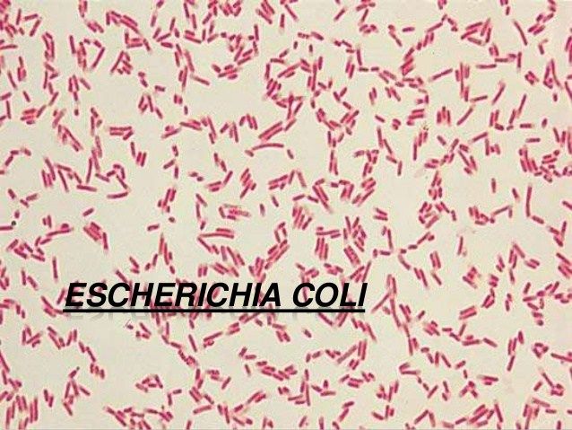
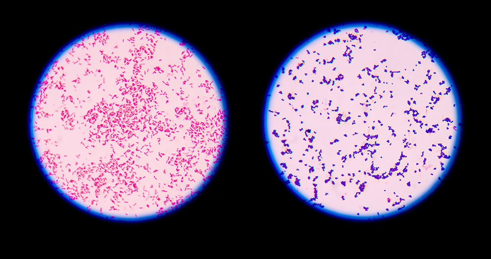
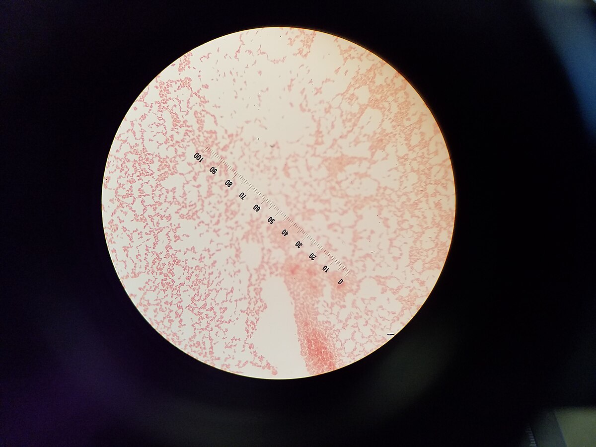
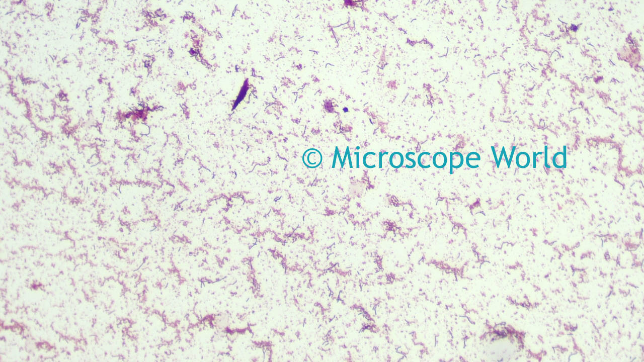
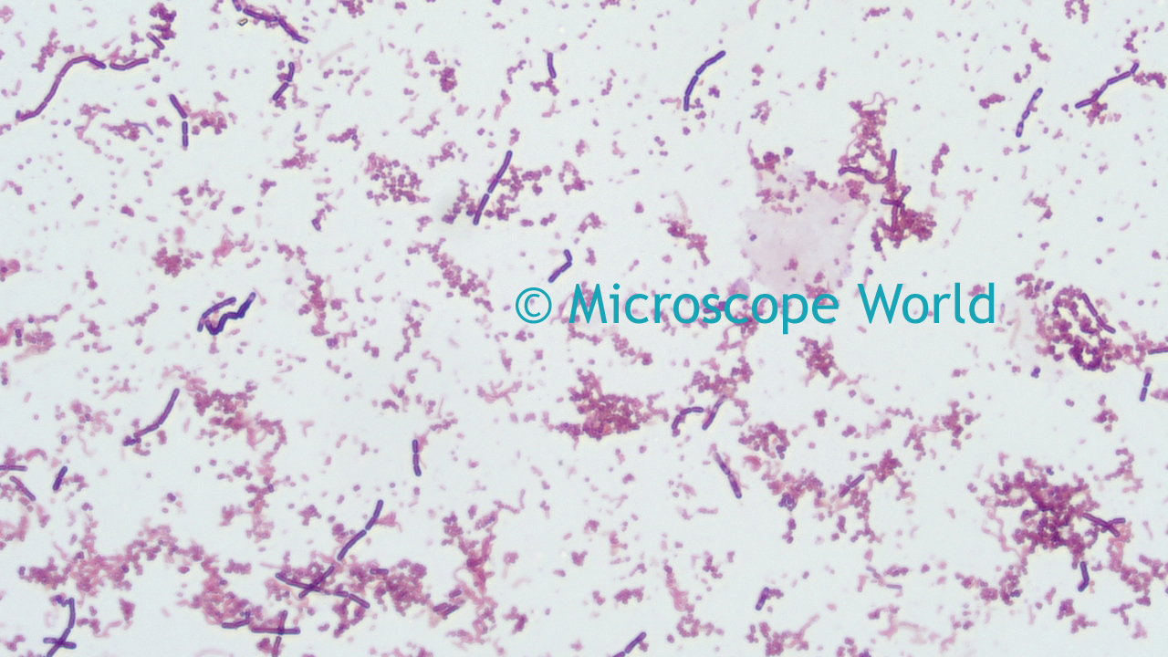
When viewed under the microscope, Gramnegative E Coli will appear pink in color The absence of this (of purple color) is indicative of Grampositive bacteria and the absence of Gramnegative E Coli Escherichia coli under 10х90х magnification using fuchsine as a dye by ElNokko (Own work) CC BYSA 40 (http//creativecommonsorg/licenses/bysa/40), via Wikimedia CommonsE coli bacteria are gramnegative, so they stain pink in a gram test E coli bacteria were first identified by bacteriologist Dr Escherich in 15 Since then, more than 700 different serotypes have been identified Some of these strains are pathogenic, but most are notMixed Gram & Gram Bacteria Under Microscope (Click on image to enlarge) Gram stain mixed sample od Staphylococcus (Gram , purple cocci) & E coli , (Gram pink rods) @ 1000xTM
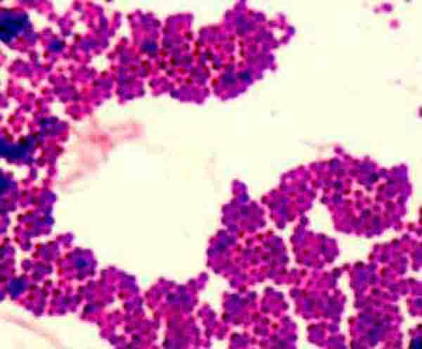


Bacterial Staining Microbiology Images Photographs And Videos Of Gram Acid Fast Endospore
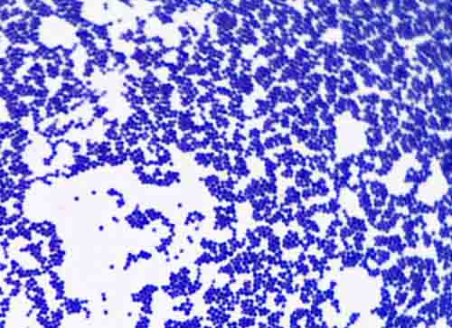


Bacterial Staining Microbiology Images Photographs And Videos Of Gram Acid Fast Endospore
Gram stain allows you to classify into Gramnegative rods (which includes E coli) and Grampositive rods (which include Bacillus, Corynebacterium, and some others)Instead, their genetic material floats uncoveredThe gram stain, originally developed in 14 by Christian Gram, is probably the most important procedure in all of microbiology It has to be one of the most repeated procedures done in any lab Gram was actually using dyes on human cells, and found that bacteria preferentially bind some dyes The Gram stain is a differential stain, as opposed to the simple stain which uses 1 dye


What Does An E Coli Bacteria Look Like Under A Microscope Quora


File Microbiology Gram Stain Jpg Wikimedia Commons
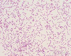
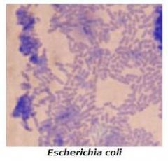
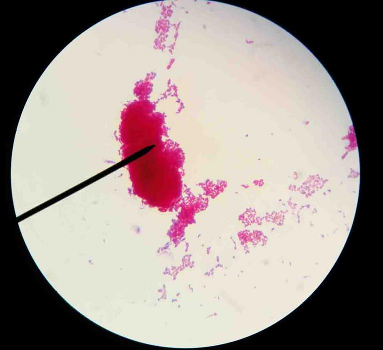
Insert figure for gram staining chemical basis here figure # Title, reference Describe the steps and what happens chemically at each step At the completion of the gram staining process, slides were dried, each smear lightly coated with a drop of immersion oil, and observed under a brightfield light microscope under the 100x objective lensE coli is a Gramnegative rodshaped bacteria When Gram stained, the organism looks pink or red Here are a couple of pictures of a Gram stain of E coli that I did under the 100X objective lens on a standard light microscope You can of course see a lot more detail under an electron microscopeA Betahemolytic colonies of Staphylococcus aureus on sheep blood agar Cultivation 24 hours, aerobic atmosphere, 37°C B Yellow colored colonies of Staphylococcus aureus on Tryptic Soy Agar Carotenoid pigment staphyloxanthin is responsible for the characteristic golden colour of S aureus colonies This pigment acts as a virulence factor


E Coli Gram Stain Page 1 Line 17qq Com


Lab 1
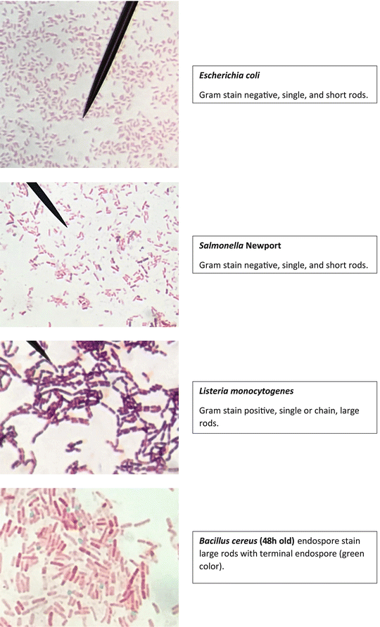
Figure 314 Gram stains Gram stains of demonstration species Below are shown typical Gram stain reactions of two species E coli (A), B cereus old (B), B cereus young (C) The images are slightly larger than what would be visible in a light microscope to improve clarityGram Staining of Micrococcus luteus,Escherichia coli, and Serratia marcescens INTRODUCTION In 14, a Danish botanist named Hans Christian Gram developed the popular staining technique known as the "gram stain" His background in studying plants at the University of Copenhagen introduced him to observing different tissues under a microscope, and thus propelled his interest in pharmacologyGram stain e coli under microscope 100x E Coli Under Microscope Gram Stain Written By MacPride Friday, June 29, 18 Add Comment Edit E Coli Gram Stain Gram Stain Images Microbiology Wayne State E Coli Bacteria Gram Negative Bacilli Gram Stain Lm X400


Pin On E Coli


What Does An E Coli Bacteria Look Like Under A Microscope Quora
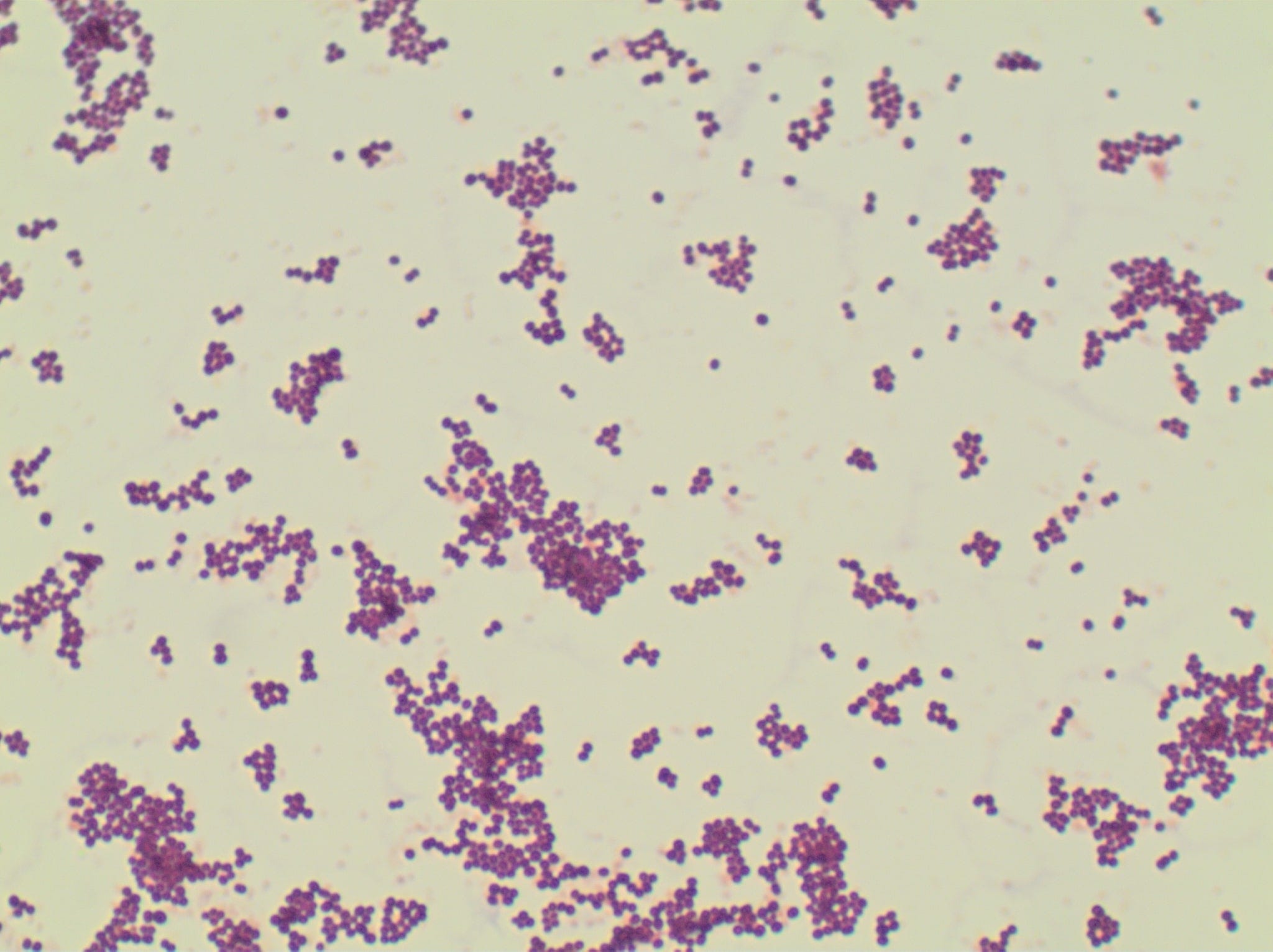
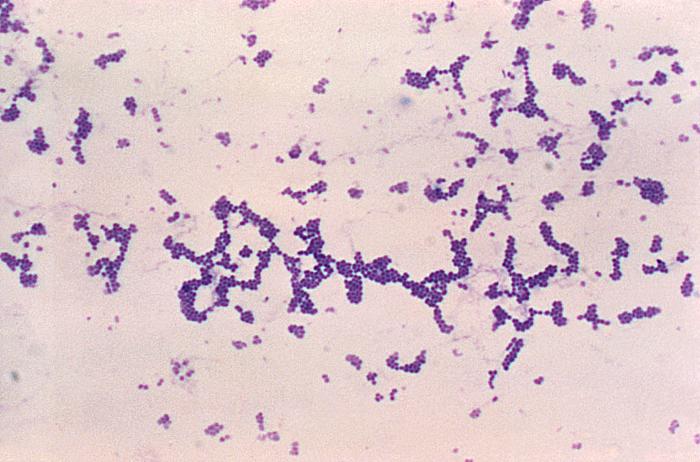
The technician decides to make a Gram stain of the specimen This technique is commonly used as an early step in identifying pathogenic bacteria After completing the Gram stain procedure, the technician views the slide under the brightfield microscope and sees purple, grapelike clusters of spherical cells (Figure 4)Show More Purpose Statement The purpose of this experiment was to determine the Gram stain of the certain slant cultures we used in class, Escherichia coli and Staphylococcus aureus, under a microscope Since it is the most important staining procedure in microbiology, it is used to distinguish between grampositive and gramnegative organisms The grampositive cells stain purple, and the gramnegative stain a pinkish colorLastly, a drop of oil was added to the slide so the sample could be viewed under the microscope using the oil immersion lens Results The E coli had a Gram Stain reaction color of pink and classified as Gramnegative Both the S epidermidis and B subtilis had a Gram Stain reaction color of purple and then classified as Grampositive (Table 1)


Tech Tip Imaging Bacteria Using Agarose Pads Biotium


E Coli Bacteria Under Microscope Page 1 Line 17qq Com
The technician decides to make a Gram stain of the specimen This technique is commonly used as an early step in identifying pathogenic bacteria After completing the Gram stain procedure, the technician views the slide under the brightfield microscope and sees purple, grapelike clusters of spherical cells (Figure 235)Bacteria species E coli and S aureus under the microscope with different magnifications Bacteria are among the smallest, simplest and most ancient livingEscherichia coli Gram stained smear under microscopeRod shapedpink in colorthat's why Gram negative Bacilli#GramStain#GNB#GNR


Gram Staining Principle Procedure And Results Learn Microbiology Online


Photomicrographs
Examples of Bacteria Under the Microscope Escherichia coli Escherichia coli (Ecoli) is a common gramnegative bacterial species that is often one of the first ones to be observed by students Most strains of Ecoli are harmless to humans, but some are pathogens and are responsible for gastrointestinal infections They are a bacillus shaped bacteria that has a very fast growth (they can double every minutes), which is one of the main reasons they are used in researchInsert figure for gram staining chemical basis here figure # Title, reference Describe the steps and what happens chemically at each step At the completion of the gram staining process, slides were dried, each smear lightly coated with a drop of immersion oil, and observed under a brightfield light microscope under the 100x objective lensE coli are Gramnegative bacteria, meaning that they do not retain the crystal violet stain commonly used to differentiate bacteria Their status as Gramnegative bacteria is due to their thin cell walls E coli has cell walls made out of two thing peptidoglycan layers, an inner and outer membrane


Solved 3 A Mixed Smear Of A 3 Day 72 Hour Culture Of S Chegg Com


Gram Negative Bacteria Images Photos Of Escherichia Coli Salmonella Enterobacter
Objective 2 To conduct a spore stain on the Bacillus culture and observe it under the microscope Results Eschericia coli Conducted a Gramstain and observed the cells under the light microscope with the 100x objective lens, giving both pink and purple rodshaped cells as shown in figure 1 Figure 1 Purple and pink E coli cells observedE coli is a Gramnegative rodshaped bacteria When Gram stained, the organism looks pink or red Here are a couple of pictures of a Gram stain of E coli that I did under the 100X objective lens on a standard light microscope You can of courseIf you were performing a Gram stain and accidentally performed the decolorization step using water rather than 95% ethanol, what would your negative control look like (under the microscope, after completion of the entire GramStain procedure)?


Laboratory Perspective Of Gram Staining And Its Significance In Investigations Of Infectious Diseases Thairu Y Nasir Ia Usman Y Sub Saharan Afr J Med


Laboratory Test 1 Flashcards Chegg Com
View under Microscope (10x, add oil then 100X) Ecoli TSA 7 Heat slide and label 8 Preform Aseptic technique to retrieve bacteria from the slant and smear on slide (remember to add a drop of water) 9 Air Dry Slide 10 Heat Fix slide 11 Preform Gram Stain 12 View under Microscope (10x, add oil then 100X) Mixed TSA and TSB 13THE PROCEDURE done individually We have cultures of E coli and Bacillus for you to gram stainThis will give you gram and gram controls to check your procedure against You can use 2 slides, 1 for each bacterium, or you can divide one slide in half and smear each bacterium on the divided slideGram Staining Now that the slide has been heat fixed, you may now begin to stain the organism This is started by applying a few drops of crystal violet for 30 seconds, then rinsing with water for 5 seconds Next, cover the slide with Gram's Iodine and let sit for a full minute before rinsing with water for another 5 seconds


Staining Introduction Types Gram Stain Afb Stain In Details


Microscopy And Staining
Insert figure for gram staining chemical basis here figure # Title, reference Describe the steps and what happens chemically at each step At the completion of the gram staining process, slides were dried, each smear lightly coated with a drop of immersion oil, and observed under a brightfield light microscope under the 100x objective lensPerform the Gram stain procedure and note the Gram reaction and cellular shape Record your results Fill out and turn in your description of your unkonwn Save your slide until your graded unknown is returned Period 4 Materials For the capsule stain 3648 hour culture of Klebsiella pneumoniae growing on a slant of EMB Agar (a highsugar medium)Gram stained smear, 100X (oil immersion) We and our partners process personal data such as IP Address, Unique ID, browsing data for Use precise geolocation data Actively scan device characteristics for identification Some partners do not ask for your consent to process your data, instead, they rely on their legitimate business interest


Www Lycoming Edu Schemata Pdfs Fritz Bacterial growth Fall14 Pdf


How E Coli Bacteria Look Like


Jcdr Bacterial Cultures Dark Field Microscopy Gram Staining Viability


E Coli Gram Stain Introduction Principle Procedure And Result Interpret


Gram Stain Images Microbiology Stain Study Tools


Photomicrographs


What Does An E Coli Bacteria Look Like Under A Microscope Quora


Gram Stain Microbiology Images Photographs


Microbe Classification Using Deep Learning By Steven Towards Data Science


Escherichia Coli Colony Morphology And Microscopic Appearance Basic Characteristic And Tests For Identification Of E Coli Bacteria Images Of Escherichia Coli Antibiotic Treatment Of E Coli Infections


Bacillus Subtilis Gram Stain Microbiology Bacillus Medical Laboratory Science


Microscope World Blog June 15


E Coli Gram Stain Page 1 Line 17qq Com


Bacterial Characteristics Gram Staining Video Khan Academy


Observing Bacteria Under The Light Microscope Microbehunter Microscopy


File E Coli Gram Stain Jpg Wikimedia Commons


Gram Stain Staphylococcus Aureus E Coli Combined In Same Organ Control Histology Slides


44 Micro Ideas Microbiology Medical Laboratory Microbiology Lab


Staphylococcus Aureus And Ecoli Under Microscope Microscopy Of Gram Positive Cocci And Gram Negative Bacilli Morphology And Microscopic Appearance Of Staphylococcus Aureus And E Coli S Aureus Gram Stain And Colony Morphology On Agar Clinical


Staphylococcus Aureus And Ecoli Under Microscope Microscopy Of Gram Positive Cocci And Gram Negative Bacilli Morphology And Microscopic Appearance Of Staphylococcus Aureus And E Coli S Aureus Gram Stain And Colony Morphology On Agar Clinical


Chapter 3 Tools Of The Laboratory E Mail


E Coli Under The Microscope Types Techniques Gram Stain Hanging Drop Method


Microbiology Lab Exercise 6 Acid Fast Staining Flashcards Cram Com


Differential Staining Gram Staining Endospore Staining Capsule Staining Foodelphi Com


Q Tbn And9gcqkye60ou Johpr02n Mbv1fferrjpdh Lnct7ymdf5qhyia1ld Usqp Cau


Lab 1 Principles And Use Of Microscope Ibg 102 Lab Reports


Gram Stain Wikipedia


56 Questions With Answers In Gram Staining Scientific Method


Bacterial Characteristics Gram Staining Video Khan Academy


Http Coltonanderson1 Weebly Com Uploads 2 4 3 0 Manual Pdf


Pin On Budi Zdrav Produzi Zivot


Gram Stain


Gram Stain


Gram Stain


Q Tbn And9gcswouuht13c4cxzkdgvwicdnmwhgkfjwlh40a Eerzzlpwjtmyt Usqp Cau


Microscopic Field Oil Immersion Objective Showing The Accumula Tion Download Scientific Diagram


Escherichia Coli Bacteria E Coli Stock Footage Video 100 Royalty Free Shutterstock


9 Gram Staining Best Practices Microbiologics Blog


Gram Stain Introduction Principle Procedure Result And Interpretation


17 Gram Stain Biology Libretexts


Staphylococcus Aureus Under Microscope Microscopy Of Gram Positive Cocci Morphology And Microscopic Appearance Of Staphylococcus Aureus S Aureus Gram Stain And Colony Morphology On Agar Clinical Significance


What Does An E Coli Bacteria Look Like Under A Microscope Quora


Solved 3 A Mixed Smear Of A 3 Day 72 Hour Culture Of S Chegg Com


Staphylococcus Aureus And Ecoli Under Microscope Microscopy Of Gram Positive Cocci And Gram Negative Bacilli Morphology And Microscopic Appearance Of Staphylococcus Aureus And E Coli S Aureus Gram Stain And Colony Morphology On Agar Clinical


What Does An E Coli Bacteria Look Like Under A Microscope Quora


Escherichia Coli Light Microscopy


Staining Technology And Bright Field Microscope Use Springerlink


Bacterial Staining Microbiology Images Photographs And Videos Of Gram Acid Fast Endospore


Staphylococcus Sp Simple Stain Methylene Blue Microbiology Microbiology Lab Bacteria Shapes


Http Faculty Fiu Edu Gantarm Ex2gramstain Pdf


Jaypeedigital Ebook Reader


Acid Fast Stain Free Microbiology Images From Science Prof Online


Details Public Health Image Library Phil


Gram Staining Principle Procedure And Results Learn Microbiology Online


Lab 1


Morphological View Of Lactococcus Culture Under Microscope 100x After Download Scientific Diagram


File E Coli Gram Stain Jpg Wikimedia Commons


Gram S Staining Of S Aureus 100x Grapes Like Black Arrow Gram Download Scientific Diagram


Gram S Staining Of S Aureus 100x Grapes Like Black Arrow Gram Download Scientific Diagram


Bacteria Under The Microscope E Coli And S Aureus Youtube


Www Banglajol Info Index Php Bjvm Article View


Vibrio Cholerae In Gram Stain Introduction Procedure And Result Interpret


Microscope World Blog June 15


Details Public Health Image Library Phil


Www Mccc Edu Hilkerd Documents Bio1lab3 Exp 4 Pdf


Gram Staining And Using A Microscope


Microscope World Blog June 15


What Does An E Coli Bacteria Look Like Under A Microscope Quora
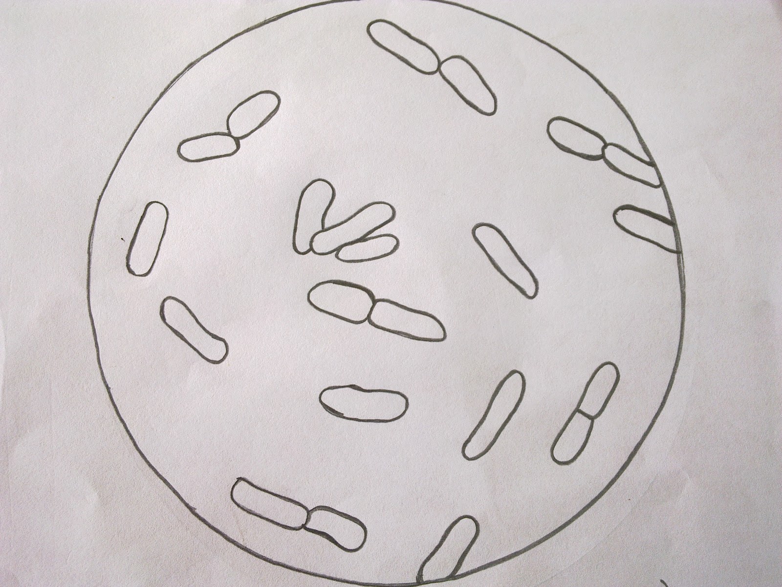


Zkfaa Bioproses Lab 1 Principles And Use Of Microscope


5 Steps Of Gram Staining Procedure How To Interpret The Results New Health Advisor
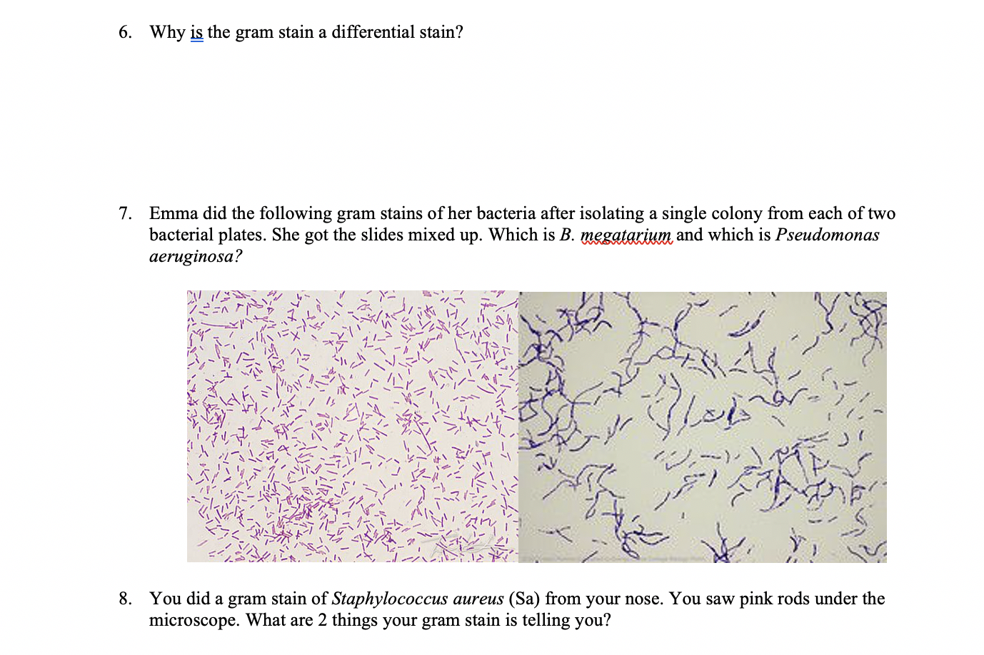


Solved 3 You Did A Gram Stain But Are Having Trouble See Chegg Com


Q Tbn And9gcqkye60ou Johpr02n Mbv1fferrjpdh Lnct7ymdf5qhyia1ld Usqp Cau


Microscopy And Staining
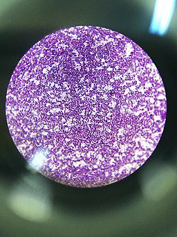


Gram Positive And Gram Negative Bacteria Structures Characterisitics


Gut Bacteria Escherichia Coli Under Microscope Youtube


Observing Bacteria Under The Light Microscope Microbehunter Microscopy
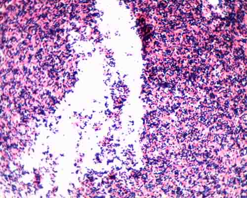


Gram Stain Microbiology Images Photographs


Gram Negative Spirillum W M Gram Stain Microscope Slide Science Lab Microbiology Supplies Amazon Com Industrial Scientific


コメント
コメントを投稿